Eye Health

See what we can see

Good eye care goes beyond a prescription. Advanced imaging lets us look closely at the health of your eyes and keep a record we can compare at every visit.

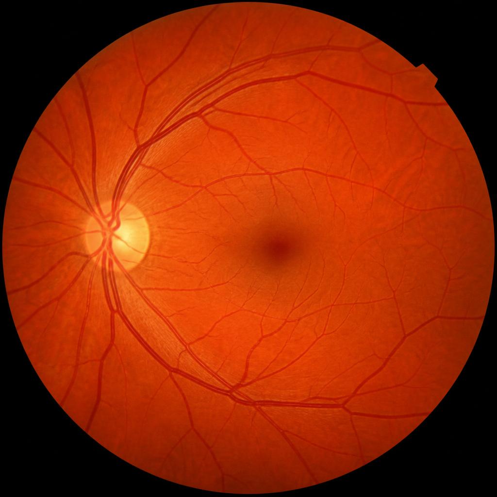
Illustrative fundus photograph of a retina showing the optic disc and blood vessels

Fundus Photography

A detailed photograph of the retina

Fundus photography captures a detailed image of the back of the eye, helping us monitor the retina, the optic nerve and the blood vessels. It is quick, comfortable and gives us a record to compare over the years.

Images for illustration purposes.
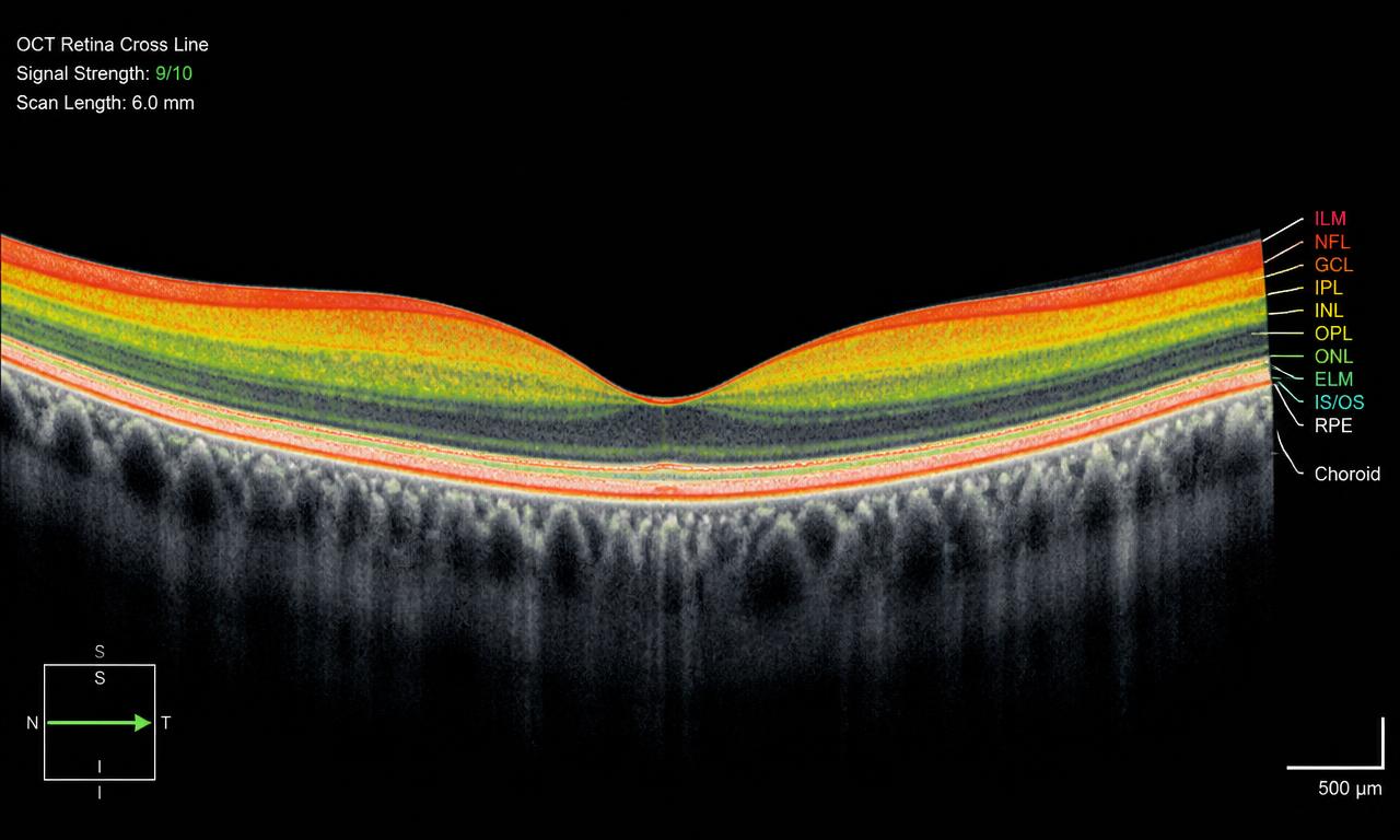
Illustrative OCT scan showing cross-sectional layers of the retina

OCT — Optical Coherence Tomography

Cross-sections of the retina

An OCT scan creates detailed cross-sectional images of the retina, which help us assess eye health and monitor changes over time.

Images for illustration purposes.

What We Look At

Part of every eye health assessment

Retinal health

The retina lines the back of the eye. Imaging lets us record its appearance and compare it over time.

Optic nerve

The optic nerve carries what you see to the brain. Its appearance is part of every eye health check.

Blood vessels

The eye is the one place blood vessels can be viewed directly — useful in diabetes and blood pressure monitoring.

Eye pressure

Measuring pressure inside the eye forms part of routine eye health screening.

This information is general and educational. It does not constitute a diagnosis, and no particular outcome is implied or guaranteed. Please book an examination for advice specific to your eyes.